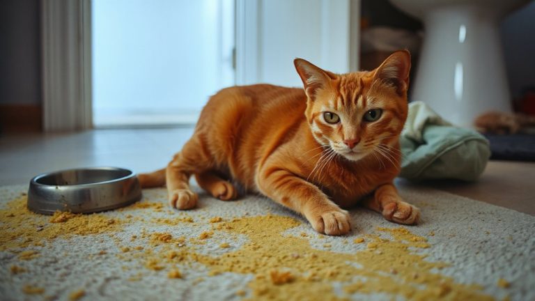
How Veterinarians Evaluate Joint Flexibility in Arthritic Pets
Your vet assesses joint flexibility by manually moving each joint through its full range of motion, noting resistance or pain. They use goniometry to measure flexion and extension angles, comparing results to breed-specific norms; deviations over 15 degrees signal significant compromise. Asymmetry, crepitus, or reduced arc indicate arthritis. Gait analysis and force plate measurements detect weight-bearing imbalances exceeding 20%. These objective tools help track disease progression and guide treatment. You’ll discover how home observations align with clinical findings.
Notable Insights
- Veterinarians assess joint flexibility using goniometry to measure range of motion and detect deviations beyond 15 degrees from breed norms.
- Hands-on manipulation evaluates joint flexion and extension, noting pain, resistance, or asymmetry during movement.
- Gait analysis observes walking, trotting, and turning to identify lameness, head bobbing, or limb asymmetry greater than 15%.
- Force plate analysis quantifies weight-bearing deficits, detecting imbalances when affected limbs bear more than 20% less force.
- Joint crepitus and muscle atrophy are assessed palpably and visually to support diagnosis of cartilage degeneration and disuse.
Why Joint Flexibility Matters for Arthritic Pets
While your pet may still move around, reduced joint flexibility often signals advancing arthritis. Maintaining joint health is critical, as stiff joints limit range of motion, impair mobility, and accelerate cartilage degradation. You’ll notice decreased activity tolerance-your dog may struggle with stairs or jump less. Veterinarians measure joint flexion and extension using goniometry, comparing results to breed-specific norms; deviations beyond 15 degrees indicate significant compromise. Reduced flexibility directly impacts pain management, increasing inflammatory mediators and muscle atrophy. Synovial fluid viscosity declines, diminishing natural lubrication. Interventions like controlled exercise, NSAIDs, and polysulfated glycosaminoglycans help preserve motion. Physical therapy protocols often include passive range-of-motion exercises performed two to three times daily for 10-minute intervals. Joint flexibility isn’t just comfort-it’s a measurable component of long-term function. Prioritizing it slows disease progression and improves clinical outcomes.
Signs of Joint Pain You Can Spot at Home
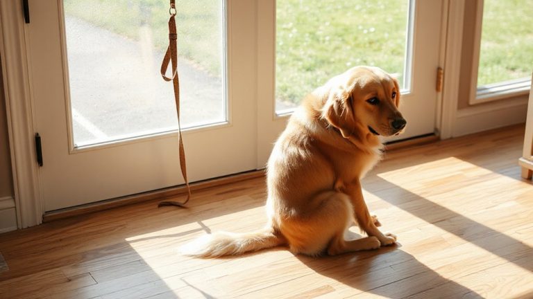
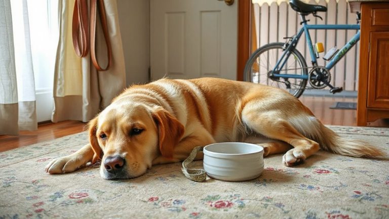
You can detect joint pain in your pet by observing subtle changes in movement and behavior. Limping behavior, even when intermittent, often indicates discomfort during weight-bearing. A common sign is reluctance to jump, especially onto furniture or into vehicles. Stiffness after rest, reduced activity, and altered gait patterns also suggest joint issues.
| Sign | Description | Frequency to Note |
|---|---|---|
| Limping behavior | Uneven stride or favoring a limb | During or after activity |
| Reluctance to jump | Avoids heights once easily managed | Consistent avoidance |
| Stiff walking | Difficulty rising or slow initial movement | Morning or post-rest |
| Licking joints | Localized grooming due to discomfort | Daily or prolonged |
| Muscle atrophy | Thinning limbs from reduced use | Visible over weeks |
Monitor these signs over time for patterns.
How Vets Test Joint Mobility and Stiffness
When evaluating joint mobility and stiffness, veterinarians follow a standardized hands-on evaluation to detect limitations in range of motion and signs of discomfort. They flex and extend each joint through its full arc, noting resistance, pain response, or asymmetry. Reduced motion often signals capsular fibrosis or osteophyte formation. Joint auscultation may reveal crepitus-grating sounds indicating cartilage degeneration. You’ll notice subtle signs, like reluctance to move, during manipulation. Gait symmetry is assessed by observing limb use at walk and trot; asymmetries suggest compensatory movement due to pain. Force plate analysis can quantify weight distribution imbalance, often showing >20% reduction in load on affected limbs. The veterinarian compares contralateral joints to identify mild deficits. Palpation includes temperature checks-increased heat implies active inflammation. These objective findings guide treatment intensity. Accuracy depends on consistent technique and patient relaxation.
Common Movement Exams for Arthritis
Veterinarians rely on standardized movement exams to assess the functional impact of arthritis on a pet’s daily mobility. You’ll often see your vet observe your pet walking, trotting, and turning in straight lines and circles. These movements allow for gait analysis, which evaluates stride length, joint excursion, and weight distribution. You should note any limping, head bobbing, or uneven pacing. Limb symmetry is measured by comparing joint angles and stance duration between left and right limbs. Asymmetry greater than 15% often indicates pain or dysfunction. High-speed video or force plates may quantify weight-bearing differences. The vet might ask you to record home videos, since stiffness can improve with warming up. These exams detect subtle changes before they become severe. You’ll get objective data from consistent, repeatable tests. They’re essential for tracking progression without guesswork.
Building a Treatment Plan for Pet Joint Pain
Though arthritis can’t be reversed, effective treatment plans slow progression and greatly reduce discomfort. Your vet will design a multimodal approach targeting inflammation, mobility, and joint health. Weight management is critical-every excess pound increases joint stress and accelerates cartilage wear. A 10% reduction in body weight can decrease lameness by up to 30% in overweight dogs. Laser therapy reduces inflammation and stimulates cellular repair; Class IV lasers deliver wavelengths of 800–905 nm at power outputs of 5–15 watts, penetrating deep into joint tissues. Treatments last 5–10 minutes and are typically administered 2–3 times weekly for 3–4 weeks. Your pet may also receive NSAIDs, joint supplements like glucosamine and chondroitin, or injectable polysulfated glycosaminoglycans. Exercise modification supports joint function without overloading compromised structures. Consistent monitoring guarantees plan effectiveness and allows timely adjustments.
On a final note
You can monitor your pet’s joint health by observing daily movements. Reduced range of motion indicates stiffness. Vets measure this using goniometry, recording joint angles to the nearest degree. A decrease of 15–20° in flexion or extension suggests significant arthritis. Treatment combines NSAIDs, weight management, and physical therapy. Motion analysis and force-plate gait studies may guide adjustments. Regular re-evaluation guarantees ideal function. Early, precise intervention preserves mobility and slows degeneration.