How Veterinarians Assess Hydration Status in Cats and Dogs
You can assess your pet’s hydration by checking their gums, which should be moist and pink; sticky or pale gums suggest fluid loss. Perform a skin pinch test: slow recoil over the shoulders indicates dehydration-under 1 second is normal, over 2 seconds signals severe fluid deficit. Sunken eyes often appear after 6–8% fluid loss. Monitor capillary refill time and heart rate, as tachycardia and delays beyond 3 seconds reflect poor perfusion. Blood tests revealing elevated PCV, BUN, and electrolyte imbalances confirm hidden deficits. Further clinical signs and treatment options follow.
Notable Insights
- Veterinarians assess hydration by checking gum moisture, as dry or sticky gums indicate dehydration.
- Skin elasticity is tested via the skin pinch test, with slow recoil suggesting fluid deficit.
- Sunken eyes and dry conjunctiva often signal moderate to severe dehydration in cats and dogs.
- Capillary refill time longer than 2 seconds may indicate poor perfusion and dehydration.
- Blood tests revealing elevated PCV, BUN, and creatinine help confirm dehydration and organ impact.
How Dehydration Damages Pets’ Organs
Water is the foundation of cellular function, and when your pet becomes dehydrated, every organ system begins to suffer. Electrolyte imbalance disrupts nerve signaling and muscle contractions, impairing heart and respiratory function. Sodium, potassium, and chloride levels shift dangerously, reducing cellular osmolarity and compromising nutrient transport. Without adequate fluid volume, blood pressure drops, decreasing perfusion to essential organs. This causes organ stress, particularly in the kidneys and liver, which rely on consistent blood flow for filtration. Metabolic waste accumulates as glomerular filtration rates decline, increasing blood urea nitrogen and creatinine. Persistent dehydration forces organs to work harder with diminished resources, like an engine running without proper coolant. Cellular metabolism slows, reducing energy production. Left untreated, ischemic injury can lead to acute organ failure. Even mild dehydration-just 5% fluid loss-alters physiological stability. Rehydration must be gradual to avoid cerebral edema.
Checking Gums: Sticky or Pale?
Your pet’s gums offer a real-time snapshot of hydration status and circulatory health. Check the moisture level by gently lifting the lip and touching the gums. Well-hydrated pets have slick, moist gums that feel like wet leather. If they feel sticky or dry, dehydration is likely present. Gum color is equally telling. Press a finger against the gum to blanch it, then release-capillary refill time should be under two seconds. Healthy gum color ranges from light pink to salmon, depending on pigmentation. Pale, white, blue, or yellowish gums signal problems: anemia, shock, or liver issues. Abnormal gum color combined with tacky moisture level indicates urgent concern. This assessment requires no tools but delivers critical data quickly. It reflects blood perfusion and fluid balance simultaneously. Always interpret gum findings alongside other clinical signs for accuracy.
Skin Pinch Test: Slow Recoil = Trouble
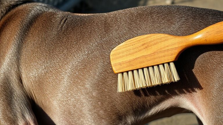
Pinching the skin over your pet’s shoulders is a standard method vets use to assess hydration. This technique evaluates skin elasticity, which reflects underlying hydration levels. Normally, the skin snaps back quickly. Delayed recoil suggests dehydration. The slower the return, the more severe the fluid deficit.
| Recoil Time | Hydration Status | Clinical Implication |
|---|---|---|
| <1 second | Normal | Skin elasticity intact |
| 1–2 seconds | Mild dehydration | ~5-6% fluid loss |
| >2 seconds | Severe dehydration | ~8-10% fluid loss |
Use your thumb and forefinger to lift a tent of skin. Release gently. Observe the return speed. In older animals, reduced skin elasticity may occur naturally, so interpretation must consider age. Consistent technique guarantees accurate assessment. Always correlate findings with other clinical signs.
Sunken Eyes? A Red Flag for Dehydration
Why might your pet’s eyes appear recessed, as if pulled inward? Sunken eyes signal severe dehydration. When fluid loss exceeds 6–8% of body weight, orbital fat and tissue volume decrease, causing eyes to retract. This is especially noticeable in dogs and cats with large, prominent eyes. You might also observe dull corneas or dry conjunctiva. Eye discharge can worsen due to reduced tear production, increasing irritation and infection risk. Vision changes may occur as dehydration affects ocular pressure and retinal perfusion. The globe’s position diminishes turgor, resembling a deflated balloon losing internal support. Unlike mild dehydration, sunken eyes indicate advanced fluid deficit requiring immediate veterinary assessment. Do not mistake this for age-related enophthalmos. Accompanying symptoms like lethargy or dry gums confirm the urgency. This clinical sign, combined with poor skin elasticity, demands rapid intervention. Fluid therapy is often necessary to restore hydration and prevent systemic complications.
Heart Rate and Capillary Refill: Vital Clues
When evaluating hydration, changes in heart rate and capillary refill time provide critical, measurable indicators of circulatory status. You’ll notice an elevated heart rate even at rest-tachycardia-when dehydration reduces blood volume. A dog’s normal resting heart rate ranges from 60–140 bpm, while cats average 140–220 bpm; significant deviations suggest compromised perfusion. Assess heart rhythm carefully, as irregularities may indicate advanced dehydration or electrolyte imbalances. Capillary refill time (CRT) is tested by pressing on the gums until they blanch, then timing how long it takes for color to return. Normal CRT is under 2 seconds; over 3 seconds signals poor circulation. Prolonged CRT correlates with reduced venous return. Vein visibility may also diminish due to low blood volume, making jugular veins less prominent. These objective signs help you quantify dehydration severity quickly and accurately.
Blood Tests: Confirming Hidden Dehydration
A blood panel is your best tool for uncovering dehydration that isn’t obvious on physical exam. Blood tests reveal changes in concentration, such as elevated packed cell volume (PCV) and total protein, which signal fluid loss. You’ll also see increased blood urea nitrogen (BUN) and creatinine-key markers of kidney function. When dehydration reduces blood flow to the kidneys, waste products accumulate. Electrolyte imbalance is common, especially shifts in sodium, potassium, and chloride levels. These imbalances affect nerve and muscle function and must be corrected carefully. The blood chemistry profile gives you precise values, allowing targeted fluid therapy. Abnormal glucose or protein levels may also point to underlying conditions worsening dehydration. Unlike physical signs, lab results provide objective, measurable evidence. They confirm hidden dehydration even when skin elasticity and gum moisture appear normal. Monitoring these values during treatment guarantees proper rehydration.
On a final note
You must act fast when dehydration is suspected. Skin tenting over the shoulders lasting more than two seconds signals fluid loss. Pale, tacky gums suggest poor perfusion. Sunken eyes reflect severe volume depletion. Elevated heart rate and prolonged capillary refill time-over two seconds-indicate circulatory compromise. Blood tests may reveal increased packed cell volume or elevated total protein. Intravenous fluids are often required. Early detection prevents organ damage.