How Veterinarians Use Otoscopes to Diagnose Ear Infections in Pets
You rely on your vet’s otoscope to detect ear infections early. It features a rigid 3–6 mm speculum and 3x to 10x magnification, designed to navigate your pet’s L-shaped canal. A bright LED (≥500 lumens) illuminates deep structures, revealing redness, discharge, or polyps. Vets assess the tympanic membrane, cerumen texture, and signs of mites or foreign bodies. Cytology confirms bacterial rods, fungal hyphae, or allergic changes. Early detection prevents rupture and guides precise treatment-unlocking clearer answers lies in what they observe next.
Notable Insights
- Veterinarians use otoscopes to visualize the L-shaped ear canal, enabling early detection of inflammation and infection beyond the visible range.
- Otoscopes provide 3x to 15x magnification and bright lighting to identify subtle changes like redness, swelling, and early debris accumulation.
- They allow direct examination of the tympanic membrane for cloudiness, rupture, or structural abnormalities indicative of advanced infection.
- Otoscopic examination helps differentiate types of ear debris-such as dark crumbly wax (yeast) or pus (bacteria)-guiding accurate diagnosis.
- High-magnification otoscopes detect ear mites, foreign bodies, and polyps that cause irritation and mimic or lead to secondary infections.
Why Pet Ear Infections Are Often Missed by Owners
Why do so many pet ear infections go unnoticed until they become severe? The lack of symptoms in early stages prevents timely detection. You might not see obvious signs like discharge or swelling immediately. Instead, subtle behavioral changes occur-your pet may tilt their head slightly, shake it more than usual, or scratch at one ear. These actions are often dismissed as minor habits. In reality, they signal discomfort. Ear canals in dogs and cats are L-shaped, making it hard to see deep issues without magnification. Infections start deep, beyond your visual reach. Early inflammation remains hidden, progressing silently. By the time overt symptoms appear, damage may already be present. Regular checks help, but without proper tools, you’re limited. Behavioral changes are often the only clue you have-don’t ignore them. Early recognition improves treatment outcomes markedly. Using a high-quality dog ear cleaner can help maintain ear health between veterinary visits.
How Otoscopes Reveal Hidden Ear Problems in Pets
How do veterinarians see what you can’t? They use otoscopes-precision tools designed to examine your pet’s ear anatomy in detail. The ear canal in dogs and cats is long and L-shaped, making visual inspection impossible without magnification. Otoscopes feature a light source and a 3x to 10x magnifying lens, allowing vets to view deep into the horizontal and vertical canals. You might notice head shaking, but only an otoscope reveals issues like polyps, trapped debris, or structural abnormalities. Wax buildup often hides beneath the surface, and when compacted, it can mimic more serious conditions. With a rigid speculum typically ranging from 3 to 6 mm in diameter, the otoscope gently enters the canal without compression. This direct visualization detects early-stage obstructions and anatomical irregularities before they become painful. Early detection means faster, more accurate treatment-all because the vet sees what you can’t. Regular cleaning with a veterinarian-approved best dog ear cleaner can help prevent many of these issues.
What Vets See During an Ear Infection Exam
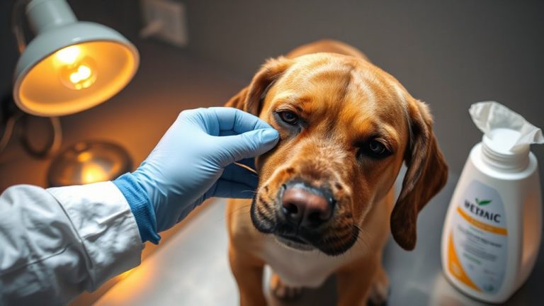
What does your pet’s ear look like when an infection takes hold? Using an otoscope, your vet examines ear anatomy closely, spotting redness, swelling, and discharge in the ear canal. In early infection stages, you might see mild inflammation and slight wax buildup. As the infection progresses, the canal narrows due to swelling, making debris harder to see without magnification. The tympanic membrane may appear cloudy or ruptured in advanced cases. Veterinarians assess cerumen color, odor, and consistency-dark, crumbly matter often suggests yeast, while pus points to bacterial involvement. Otoscopic lenses provide 6x to 15x magnification, enabling clear visualization deep in the horizontal canal. Precise lighting isolates abnormalities like ulcers, polyps, or foreign bodies. Early detection supports timely treatment. Recognizing changes across infection stages guarantees accurate diagnosis and prevents chronic damage to delicate ear structures.
Bacterial vs. Fungal vs. Allergy-Related Ear Infections
While some ear infections in pets look similar on the surface, their underlying causes-bacterial, fungal, or allergy-related-require different treatments and can be distinguished with otoscopic evaluation and cytology. You’ll see red, swollen ear canals with all three, but microscopic analysis reveals the real culprit. Bacterial overgrowth typically involves cocci or rod-shaped organisms, often linked to underlying moisture or trauma. Fungal invasion shows long, branching hyphae on cytology, commonly caused by Malassezia species thriving in warm, oily environments. Allergy-related infections lack primary pathogens but display chronic inflammation and ceruminous gland hyperplasia. Otoscopic detail-like ulcerated epithelium in bacterial cases or copious brown debris in fungal ones-guides diagnosis. You need this precision because antibiotics won’t resolve fungal issues, and antifungals won’t fix bacterial overgrowth. Misdiagnosis leads to recurrence. Accurate identification through staining and magnification guarantees targeted, effective therapy.
Ear Mites and Foreign Objects: Common Hidden Causes
Could something as small as a grain of sand be causing your pet’s constant head shaking? Yes-foreign bodies like plant material or debris often trigger irritation. Ear mites, though tiny, are another frequent culprit, especially in cats and young dogs. These parasites feed on skin oils and wax, leading to inflammation and dark, crumbly discharge. Without an otoscope, both issues can be missed during a basic ear exam. Regular cleaning with a veterinarian-recommended cat ear cleaner can help prevent buildup that exacerbates these conditions.
| Cause | Key Characteristics |
|---|---|
| Ear mites | Microscopic mites (*Otodectes cynotis*), highly contagious, cause intense itching and waxy debris |
| Foreign bodies | Grass seeds, sand, or hair; often lodged in the vertical ear canal, causing unilateral symptoms |
Veterinarians use otoscopes with magnification (5–20x) and bright LED light (≥500 lumens) to detect these hidden triggers. Accurate identification allows targeted treatment-critical for resolving discomfort and preventing secondary infections.
How Early Otoscope Use Prevents Serious Ear Damage
When you notice your pet tilting their head or scratching at one ear, an ot游戏副本scope exam early in the veterinary visit can stop minor irritation from becoming permanent damage. Early detection allows vets to identify inflammation, infection, or blockages before they worsen. Using an otoscope with 3.5x to 5x magnification and adjustable LED lighting, veterinarians clearly visualize the ear canal and tympanic membrane. This precision supports preventive care by catching issues like early-stage otitis or wax buildup. Without intervention, chronic infections can lead to calcification, hearing loss, or the need for surgery. Detecting problems at the first sign means treatment is simpler, less invasive, and more effective. You reduce long-term risks markedly. Regular ear checks during routine visits enhance early detection outcomes. An otoscope’s 1.9 cm (0.75 in) speculum fits most pets comfortably, enabling thorough exams. Early action protects your pet’s auditory health and overall well-being.
On a final note
You rely on an otoscope to detect early signs of otitis in pets. Its 3–5 mm speculum accesses narrow canals. High-intensity halogen or LED lighting illuminates debris, swelling, or ulceration. Magnification (3–6×) reveals erythema, discharge consistency, or moving mites. Pneumatic function assesses tympanic membrane mobility. Immediate visualization prevents progression to chronic disease. Regular otoscopic exams reduce risk of rupture, hearing loss, or middle ear involvement. Early diagnosis means faster, targeted treatment.