How Veterinarians Assess Spinal Pain in Reluctant or Aggressive Pets
You can spot spinal pain in aggressive or reluctant pets by observing posture and movement first. Asymmetrical weight bearing, shortened stride, or a hunched back often signal discomfort. High-resolution motion analysis at 120 frames per second detects subtle gait changes, while infrared thermal imaging identifies inflammation through temperature differences as small as 0.1°C along the spine. Behavioral cues like tail flicking or ear flattening correlate with pain. When needed, sedation allows safe, accurate assessment-revealing more than behavior alone ever could.
Notable Insights
- Veterinarians use remote observation of posture, gait, and movement to detect spinal pain without physical contact.
- Infrared thermal imaging identifies inflammation or nerve dysfunction through precise temperature differences along the spine.
- High-speed motion analysis quantifies abnormalities like shortened stride or trunk instability in unrestrained animals.
- Minimal-contact techniques, including light touch and draped exams, reduce stress during spinal assessment.
- Sedation with drugs like dexmedetomidine allows safe, thorough evaluation when aggression or fear impedes examination.
How Vets Detect Spinal Pain Without Touch
While you might expect a hands-on exam to be the first step, veterinarians often begin evaluating spinal pain in pets through careful observation of posture and movement. This non invasive assessment allows clinicians to detect discomfort without triggering fear or aggression. Subtle shifts in weight distribution, head carriage, or gait asymmetry can indicate spinal dysfunction. You can observe these behaviors during voluntary activity or controlled walking tasks. Remote monitoring systems, using high-resolution video and motion-sensor technology, enhance detection accuracy. These systems record kinematic data-such as stride length, limb placement, and trunk stability-over time. Quantitative analysis identifies deviations from baseline movement patterns, often before obvious symptoms arise. Infrared thermal imaging may supplement this approach, revealing temperature asymmetries along the spine linked to inflammation. Together, these tools enable objective, repeatable assessments. They minimize handling stress while improving diagnostic precision. Remote monitoring and non invasive assessment are transforming how you detect spinal pain-early, safely, and effectively.
Body Language Clues That Reveal Back Discomfort
You can spot back discomfort in your pet by learning the specific body language signs that signal spinal distress. Tail flicking is a key indicator, often occurring when your pet shifts position or moves, reflecting irritation or pain along the lumbar or sacral regions. Ear flattening, especially when repeated or sustained, correlates with discomfort and may precede aggression in painful animals. Your pet may avoid lying on its side or hesitate to jump, revealing reduced spinal flexibility. A hunched posture suggests muscle guarding, a protective response to vertebral instability. Changes in gait, such as shortened stride length or uneven weight distribution, further support spinal involvement. These visual cues help you assess pain severity without touch. Observing frequency, duration, and context of tail flicking or ear flattening improves diagnostic accuracy. Combined, these behaviors offer objective data for early intervention.
Imaging and Tools That Avoid Physical Contact
How do veterinarians confirm spinal pain without touching your pet? They use remote sensing and thermal imaging to detect abnormalities. Thermal imaging cameras measure skin surface temperature, identifying areas of inflammation or altered blood flow along the spine. These infrared cameras detect temperature differences as small as 0.1°C. Elevated heat often indicates inflammation, while cooler regions may suggest nerve dysfunction. The data appears in color-coded maps, allowing vets to pinpoint pain zones without contact. Remote sensing tools, like motion analysis systems, track gait changes using high-speed video and reflective markers. These systems record movement at 120 frames per second, revealing subtle lameness or spinal rigidity. Combined, thermal imaging and remote sensing provide objective, non-invasive data. They’re especially useful for aggressive or fearful animals. You get accurate diagnostics while minimizing stress. These tools support early detection and ongoing monitoring, improving treatment outcomes without direct physical exams.
Gentle Handling Techniques for Reactive Pets
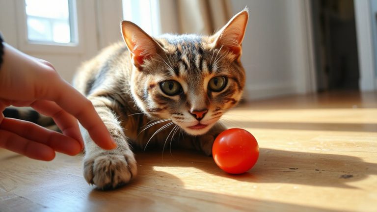
A calm, structured approach is essential when evaluating spinal pain in reactive pets. You must prioritize a calm approach to minimize stress and resistance. Begin with minimal restraint, using a soft, non-slip mat to prevent slipping. Apply light touch assessments with gloved hands, starting away from the spine to initiate trust building. Gradually progress to palpation, using 50–100 grams of pressure-equivalent to the weight needed to indent a ripe tomato. Avoid sudden movements. Use towel draping for anxious cats, ensuring only one body region is exposed at a time. Keep sessions under five minutes to reduce fatigue and fear. Tools like hemostats or blunt probes can assess withdrawal reflexes at a safe distance. Consistent verbal cues and pauses reinforce predictability. These techniques enhance cooperation without sedation, increasing diagnostic accuracy in pets exhibiting fear or pain-induced aggression.
Silent Pain Signals: What Behavior Reveals
Why might your pet not yelp, even when in pain? Animals often suppress vocal cues to avoid appearing vulnerable, especially in stressful environments. Instead of relying on cries or whimpers, observe subtle shifts in behavior. Changes in rest patterns-such as difficulty lying down, frequent shifting, or reluctance to rise-can indicate spinal discomfort. Your pet may avoid jumping, hesitate on stairs, or stand with an uneven posture, distributing weight to reduce spinal load. These actions reflect nociceptive processing, where pain alters motor function. Reduced movement or prolonged sleeping isn’t always laziness; it’s often pain avoidance. Monitor duration, frequency, and context of these behaviors. A cat sleeping 18 hours daily with rigid positioning differs from normal rest patterns. Dogs may withdraw socially or resist grooming. These silent signals are clinically significant, offering objective data points for assessment, much like essential signs in human medicine.
When Sedation Enables Accurate Diagnosis
Signs of spinal pain in pets often manifest quietly, with altered movement and posture offering the first clues. When your pet won’t sit still, resists touch, or acts aggressively, physical exams become unreliable. That’s when sedation benefits become clear. Light to moderate sedation, using agents like dexmedetomidine or butorphanol, minimizes stress and muscle tension, allowing for a thorough neurological and orthopedic assessment. This leads to improved diagnostic precision, enabling veterinarians to detect subtle spinal misalignments, localized pain responses, or neuromuscular deficits. Imaging such as radiographs or MRI often requires absolute stillness-sedation guarantees accurate image capture without motion artifacts. Dosage is weight-adjusted and species-specific, minimizing risk. Without restraint-induced distortion, veterinarians can map pain zones, assess range of motion, and identify nerve root compression. Sedation isn’t avoidance-it’s a tool for clarity, guaranteeing your pet’s diagnosis is based on objective findings, not guesswork.
On a final note
You rely on objective indicators when hands-on exams aren’t possible. Cats may crouch with arched backs; dogs avoid jumping-signs linked to thoracolumbar discomfort. Video gait analysis detects subtle lameness at 1.5–2.0 m/s walk speed. Thermal imaging shows asymmetrical surface temperatures exceeding 1.5°C, indicating inflammation. Low-field MRI achieves 0.25–0.3 Tesla resolution for disc pathology. Sedation with dexmedetomidine (5–10 μg/kg) enables safe imaging. Behavior and technology replace touch without compromising diagnostic accuracy.