How Early Detection of Elbow Dysplasia Prevents Arthritis in Dogs
You can prevent arthritis by catching elbow dysplasia early. Signs like post-exercise limping or joint swelling in dogs as young as 4–5 months warrant immediate evaluation. Radiographs under sedation or CT scans detect lesions as small as 1–2 mm, such as fragmented coronoid processes. Early surgical correction stabilizes the joint, halting cartilage degeneration. Even minor weight reductions-just 10%-decrease joint load by up to 40 pounds per step. Specific interventions at the first sign of lameness change long-term outcomes. More details on breed-specific risks and imaging protocols follow.
Notable Insights
- Early detection identifies joint abnormalities before cartilage damage progresses to irreversible arthritis.
- Prompt diagnosis allows interventions that reduce inflammation and slow degenerative joint changes.
- Radiographic and CT imaging in young dogs reveal subtle lesions before clinical arthritis develops.
- Early management with joint supplements supports cartilage health and delays arthritic onset.
- Weight control and activity modification from an early stage decrease joint stress and arthritis risk.
Signs of Elbow Dysplasia in Dogs
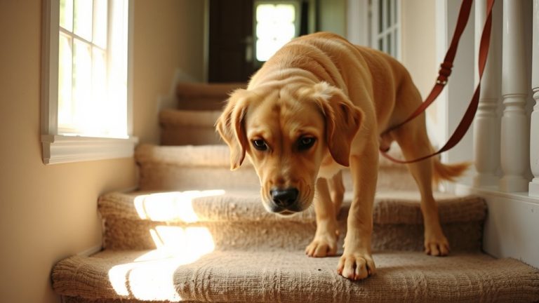
A noticeable sign of elbow dysplasia in your dog is limping, especially after exercise or when getting up from rest. You’ll often observe front limb lameness, typically worsening over time. Lameness evaluation by a veterinarian confirms asymmetric gait patterns, sometimes with a nodding head motion during stride. Joint inflammation follows abnormal cartilage wear, producing heat, swelling, and pain on palpation. Radiographic imaging reveals secondary osteoarthritis, OCD lesions, or fragmented medial coronoid process. Early diagnosis relies on dynamic assessment, including gait analysis and CT scans for precise anatomical detail. Affected dogs may carry 60–70% of their weight on the forelimbs, increasing joint stress. Persistent inflammation accelerates cartilage degradation, leading to chronic arthritis. You should seek veterinary assessment at the first sign of irregular movement. Prompt lameness evaluation enables timely intervention, reducing long-term joint damage. Managing joint inflammation early improves prognosis and maintains function, slowing progression to debilitating osteoarthritis. Detection at initial stages is critical.
Breeds Most at Risk for Elbow Dysplasia
Large and giant-breed dogs face the highest risk for developing elbow dysplasia, with genetic predisposition playing a central role. You’ll see this condition most often in breeds like Labrador Retrievers, Golden Retrievers, German Shepherds, Rottweilers, and Bernese Mountain Dogs. These breeds inherit traits that contribute to improper joint development. Genetic predisposition means abnormal growth patterns are passed down, increasing susceptibility. Conformation faults-such as incorrect angulation of the humerus, radius, or ulna-cause uneven weight distribution across the joint. This leads to abnormal wear, cartilage damage, and eventual osteoarthritis. Skeletal maturity studies show at least 20% of affected breeds exhibit radiographic signs by 12 months. Males are slightly more prone due to faster growth rates. Early screening through OFA or PennHIP evaluations helps identify subtle conformation faults before symptoms arise. You can mitigate long-term damage with timely intervention.
What Is Elbow Dysplasia? (And Why It Matters)
Elbow dysplasia isn’t just one disease-it’s a cluster of developmental conditions affecting the elbow joint in dogs, and it’s one of the leading causes of forelimb lameness in young large-breed dogs. You should know it often stems from a genetic predisposition, especially in breeds like Labs, Goldens, and German Shepherds. These inherited traits can lead to abnormal joint development. One key factor is joint laxity, where loose ligaments allow irregular movement, causing cartilage damage. Over time, this wear-and-tear leads to osteoarthritis. The condition typically arises during rapid growth phases, between 4 and 10 months of age. Misalignment of the radius and ulna bones disrupts load distribution. Early changes are microscopic but progressive. Without intervention, structural degeneration is inevitable. Understanding the role of genetics and biomechanics helps you make informed decisions. You can’t change DNA, but you can influence outcomes through screening and management. That’s why early awareness matters.
How Vets Diagnose Elbow Dysplasia
Your dog starts limping after play, and you’re left wondering what’s wrong. Your vet will first perform joint palpation, checking for pain, swelling, or restricted motion in the elbow. This hands-on exam helps identify abnormalities but isn’t definitive. For accurate diagnosis, imaging techniques are essential. X-rays are most common, taken under sedation to guarantee proper positioning-specific views like the 45° flexed mediolateral reveal joint incongruity or osteophytes. If early changes are subtle, CT scans offer 3D detail, detecting ununited anconeal processes or fragmented coronoid tips as small as 1–2 mm. MRI is less common but useful for soft tissue assessment, like cartilage damage. These imaging techniques confirm elbow dysplasia and rule out other conditions. Early use of precise diagnostic methods guarantees timely intervention before arthritis develops. Accuracy matters-misdiagnosis delays care.
Treatments That Stop Arthritis in Dogs
While early diagnosis of elbow dysplasia is critical, halting the progression to arthritis requires targeted intervention. You can slow joint deterioration with consistent use of joint supplements containing glucosamine, chondroitin, and MSM. These compounds support cartilage repair and maintain synovial fluid viscosity, reducing friction in the joint capsule. Ideal dosing is weight-dependent-typically 20 mg glucosamine per kg of body weight daily. Anti-inflammatory diets rich in omega-3 fatty acids (EPA and DHA) decrease pro-inflammatory cytokines like IL-1β and TNF-α. Look for veterinary-formulated foods with a minimum of 2% omega-3s by volume. Fish oil supplements at 100 mg EPA/DHA per 5 lbs of body weight have shown measurable reduction in joint inflammation markers. Always pair these with veterinarian-approved treatment plans using precise diagnostic imaging to track degenerative changes. One of the most effective ways to support long-term joint health is through the use of best dog joint supplements for hip dysplasia.
How Weight and Exercise Affect Elbow Dysplasia
Every extra pound your dog carries markedly increases stress on the elbow joints, accelerating cartilage wear and worsening dysplasia symptoms. Weight management is critical-reducing body weight by just 10% can decrease joint load by up to 40 pounds during movement. Excess fat also secretes inflammatory cytokines, exacerbating joint degeneration. Implement a measured feeding plan using a 3 oz measuring cup and a high-protein, low-calorie diet to achieve ideal body condition. Exercise modification supports joint health. Replace high-impact activities like jumping or sprinting with controlled leash walks of 20–30 minutes twice daily. Use non-slip mats on hardwood floors to reduce joint strain during movement. Swimming is ideal-providing resistance with zero joint impact. These adjustments slow arthritis progression, preserving mobility longer.
On a final note
You can prevent arthritis by catching elbow dysplasia early. Radiographs and CT scans detect joint incongruity, osteochondral defects, or fragmentation as small as 2 mm. Early surgical intervention corrects abnormalities before cartilage damage progresses. Weight control reduces joint load by up to 40%. Controlled exercise maintains range of motion without accelerating wear. Together, these steps markedly delay or halt arthritic degeneration in genetically predisposed breeds.