Pet Gait Assessment: How Vets Spot Lameness (0-4 Scale)
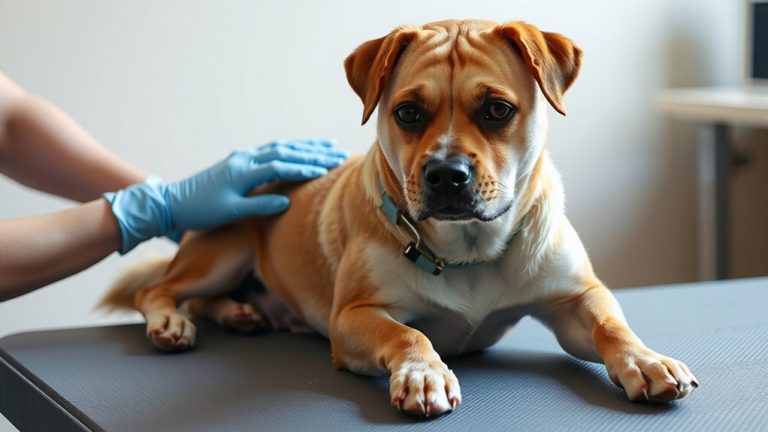
You’ll notice vets assess your pet’s gait for limb symmetry, stride length, and weight distribution. They look for a 10% or greater asymmetry in weight-bearing using force plates. Lameness scores range from 0–4, with a score of 2 indicating a visible limp at walk. Reduced joint flexion, abnormal foot placement, or head bobbing may signal pain or neurologic issues. Muscle atrophy, stiffness after rest, or altered stance phase duration provide further diagnostic clues. Patterns like hip hiking or knee buckling reveal underlying dysfunction worth further exploration.
Notable Insights
- Veterinarians assess for lameness, noting visible limps or reduced weight-bearing on affected limbs during movement.
- They evaluate weight distribution, looking for asymmetries greater than 15% that suggest pain or structural issues.
- Postural abnormalities like head tilt, head bobbing, or stiff neck are checked for potential neurologic or musculoskeletal causes.
- Gait analysis identifies deviations such as shortened stride, irregular foot placement, or joint stiffness impacting mobility.
- Muscle symmetry and joint range of motion are examined to detect atrophy, pain, or early signs of osteoarthritis.
Signs of Lameness: What to Watch in Your Pet
One of the most telling signs your pet may have a mobility issue is an obvious limp, which veterinarians classify as lameness-a deviation from a normal gait pattern. You’ll notice limping behavior when your pet bears less weight on one limb or avoids full extension during movement. This often indicates pain or structural dysfunction. Joint stiffness commonly accompanies lameness, especially after rest, and is a hallmark sign of osteoarthritis. Reduced range of motion in affected joints-measured objectively via goniometry-confirms restricted mobility. Pets may rise slowly or hesitate to jump, reflecting discomfort. Palpation often reveals crepitus or effusion in the joint capsule. Lameness scales, such as the 0–4 veterinary system, help quantify severity: a score of 2 means visible limp at a walk. Chronic cases show muscle atrophy due to disuse. Monitoring changes over time improves diagnostic accuracy. Early detection allows intervention before degeneration progresses.
How Your Dog’s Weight Shifts Reveal Pain
Your dog’s weight distribution during movement offers critical clues to underlying pain or discomfort. Uneven weight distribution often indicates an effort to off-load a painful limb. You’ll notice your dog placing less weight on the affected leg during stance phase, reducing ground contact time. This shift alters joint alignment, especially in the hips and shoulders, leading to abnormal loading patterns. Compensatory mechanics may mask the primary issue, making subtle lameness harder to detect. Veterinarians assess symmetry, measuring weight distribution across limbs using force plates or pressure-sensitive walkways when available. Deviations exceeding 15% between left and right limbs suggest pathology. Poor joint alignment during motion increases stress on ligaments and cartilage, accelerating degenerative changes. Observing gait on level surfaces helps identify these shifts early. Persistent asymmetry warrants diagnostic imaging. You can support early detection by noting reluctance to jump or rise, which reflects pain-related weight-shifting behavior.
Head Tilt or Bobbing? What Posture Says
Why does your dog tilt or bob its head while walking? These movements may signal issues with balance, pain, or neurological function. Observing ear position and neck alignment helps identify underlying causes. A consistent head tilt often reflects vestibular disease or inner ear problems, while rhythmic bobbing might suggest cervical spine instability.
| Symptom | Possible Cause | Clinical Significance |
|---|---|---|
| Head tilt | Vestibular disorder | Indicates inner ear or brainstem |
| Head bobbing | Cervical vertebral issues | Linked to neck alignment stress |
| Asymmetric ears | Ear position changes | Suggests discomfort or nerve loss |
| Stiff neck | Muscle spasms or injury | Affects gait and posture |
| Head low at rest | Chronic pain or weakness | Correlates with reduced mobility |
Abnormal posture impacts gait mechanics. Neck alignment directly influences balance and coordination. You should report persistent changes promptly.
Normal vs. Abnormal Pet Gait: Spot the Difference
How can you tell if your pet’s walk is healthy or hiding a problem? A normal gait shows proper joint alignment and consistent foot placement. Your pet’s limbs move in a balanced, rhythmic pattern. Joints bend smoothly, with no visible stiffness or overextension. Feet land squarely, with toes pointing forward and weight distributed evenly. In an abnormal gait, joint alignment is often off-hips may hike, knees buckle, or elbows twist outward. Foot placement becomes irregular, with crossing, dragging, or uneven weight-bearing. You might notice a shortened stride or a head bob during movement. These changes suggest discomfort, injury, or neurological issues. Asymmetry in limb motion or altered paw positioning can signal musculoskeletal disease. Observing your pet on level ground helps detect subtle differences. Early recognition of abnormal mechanics allows timely intervention and supports long-term joint health.
Why Vets Care About Your Pet’s Walk
A pet’s gait is more than just the way it moves-it’s a window into its neuromusculoskeletal health. You can see signs of pain or dysfunction long before your pet stops walking altogether. Veterinarians assess your pet’s stride length, foot placement, and weight distribution with precision. Subtle shifts in joint health-like decreased flexion in the stifle or radiocarpal joints-signal early osteoarthritis. Muscle tone is evaluated by symmetry and bulk, especially along the hindlimbs and back. Asymmetry or atrophy suggests disuse from discomfort or nerve issues. Lameness may not be obvious to you, but vets detect even a 10% weight-bearing asymmetry using high-speed gait analysis. Monitoring gait patterns over time reveals progression or response to therapy. It’s not just about mobility-it’s about diagnosing underlying conditions before irreversible damage occurs. Your pet’s walk tells a measurable story.
When to See a Vet for Mobility Changes
When did you last notice your pet hesitating to jump into the car or struggling to rise after resting? These subtle changes often signal underlying mobility issues. Joint stiffness commonly appears as reduced range of motion, especially after inactivity, and may indicate osteoarthritis or degenerative joint disease. You might observe your pet taking longer to stand or reluctance to climb stairs. Muscle atrophy, identified by a visible decrease in muscle mass, particularly along the limbs or spine, suggests disuse or nerve dysfunction. Asymmetry in limb girth of more than 1 cm between sides is clinically significant. Early veterinary assessment improves outcomes. Diagnostic tools like goniometry measure joint angles to quantify stiffness, while body condition scoring and muscle mass charts track atrophy progression. Radiographs or MRI may confirm structural pathology. Don’t wait-mobility changes are rarely normal aging. Timely intervention slows decline and maintains function.
On a final note
Your pet’s gait reveals critical health clues. Subtle lameness may indicate joint instability or soft tissue injury. Weight shifting reduces load on painful limbs, often seen in osteoarthritis. Head bobbing synchronizes with forelimb pain, acting like a counterbalance. Abnormal stance or stride length-below 80% of normal-signals discomfort. Vets measure range of motion; values under 120 degrees in hips suggest pathology. Early detection improves outcomes markedly.