
Vet Skin Exams: Wood’s Lamp, Cytology & Mite Scrapings Explained
Your pet’s coat should be smooth and resilient, with consistent sheen and shaft diameter-dullness or brittleness may signal nutrient deficiencies or illness. Vets inspect skin by parting fur, examining 1 cm² sections for redness, scaling, or papules. They use a Wood’s lamp at 365 nm to detect ringworm fluorescence and perform cytology at 400x magnification to identify yeast or bacteria. Skin scrapings penetrate the stratum corneum to uncover mites like Sarcoptes. Persistent flaking or itching could indicate allergies, parasites, or endocrine disorders-further evaluation reveals underlying causes.
Notable Insights
- Veterinarians assess coat condition by checking for shine, texture, and signs of thinning or excessive shedding.
- Skin is examined for redness, scaling, pigmentation changes, and lesions using visual and tactile evaluation techniques.
- Tactile exams involve parting fur to inspect small skin areas and applying pressure to detect fluid buildup.
- Diagnostic tools like Wood’s lamp, skin scrapings, and cytology swabs help identify infections or parasites.
- Persistent coat or skin issues prompt further testing for allergies, endocrine disorders, or nutritional deficiencies.
What Your Pet’s Coat Reveals About Skin Health
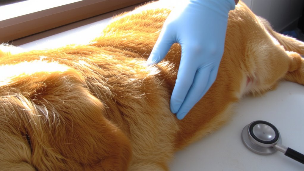
A dull or brittle coat often signals underlying skin issues. You should recognize that coat texture is a direct reflection of dermal health and overall wellness. A healthy coat feels smooth and resilient, with consistent shaft diameter and sheen. Changes in texture-such as coarseness, thinning, or excessive shedding-may indicate metabolic, hormonal, or nutritional imbalances. You might also notice pigmentation changes, including hyperpigmentation or hypopigmentation, which can point to endocrine disorders like hypothyroidism or Cushing’s disease. These shifts typically appear in areas with low sunlight exposure, such as the abdomen or inner thighs. Veterinarians assess these traits systematically, correlating them with clinical history. Monitoring coat texture and pigmentation changes helps identify issues early. You won’t always see inflammation or lesions, but subtle coat alterations provide measurable, observable indicators. Early recognition supports timely, accurate diagnosis and improves treatment outcomes.
Redness, Flaking, and Bumps: What Vets Look For
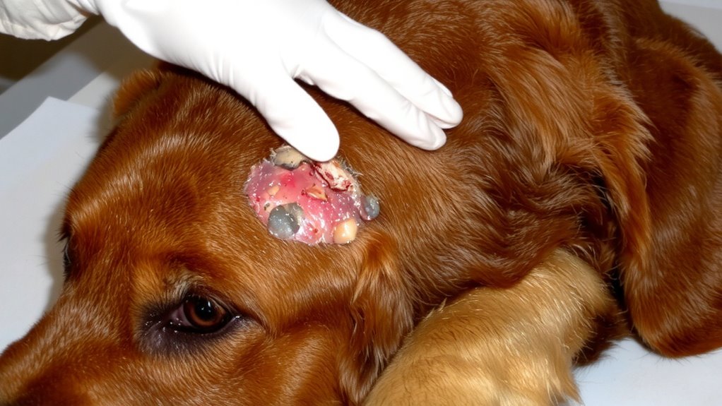
Inflammation, scaling, and papular lesions are key clinical signs vets evaluate during dermatological exams. You’ll notice redness when capillaries dilate due to irritation or infection. Vets classify this erythema by intensity and distribution-localized or generalized. Flaking, or scaling, results from accelerated epidermal turnover. Dry, bristly skin often links to poor diet quality, specifically deficiencies in omega-3 and omega-6 fatty acids. Bumps, known as papules, measure less than 1 cm in diameter and indicate possible allergic dermatitis, parasitic infestation, or infection. Vets differentiate these from pustules or nodules by size, color, and consistency. Seasonal shedding can exacerbate flaking, especially in double-coated breeds. Abnormal shedding patterns may signal underlying inflammation. Each lesion’s morphology, arrangement, and symmetry helps determine etiology. Accurate assessment requires noting onset, duration, and response to prior treatments. These observations guide diagnostic testing and management. Using a best dog shampoo for sensitive skin can help manage irritation and support the skin barrier in affected dogs.
How Vets Check for Hidden Skin Problems
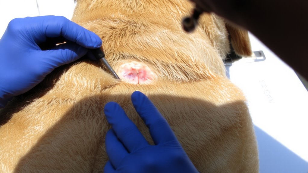
What lies beneath the surface of your pet’s skin? Vets use tactile exams and specialized tools to detect hidden lesions and underlying infections. You’ll notice them parting fur with their fingers, inspecting skin in sections no larger than 1 cm². They apply gentle pressure to check for fluctuance, a sign of fluid buildup. A Wood’s lamp helps identify fungal infections like ringworm, which fluoresce under ultraviolet light at 365 nm wavelength. Cytology swabs collect cells from the skin surface and ear canals, later examined microscopically at 400x magnification to spot bacteria or yeast. Skin scrapings penetrate the stratum corneum to detect mites such as *Sarcoptes*. If deeper issues are suspected, vets may order biopsies using 6-mm punch tools. These methods guarantee hidden lesions and underlying infections don’t go undetected. Early diagnosis improves treatment outcomes dramatically.
Causes of Itchy, Flaky, or Thin Pet Coats
Though you might first notice flaking or excessive scratching, these symptoms often signal underlying disruptions in your pet’s skin barrier and hair growth cycle. Allergies are a leading cause, triggering inflammatory responses that compromise skin integrity. Common allergens include pollen, mold, dust mites, and certain proteins in food. These activate immune pathways, increasing histamine release and causing itch. Parasites like fleas, mites, and lice directly irritate the skin, feeding on blood or burrowing into tissue. Flea saliva alone contains over 15 antigens that provoke hypersensitivity. Demodex mites disrupt follicular structure, leading to hair loss and scaling. Both allergies and parasites alter sebum production, weakening the coat’s protective lipid layer. Secondary yeast or bacterial infections often follow, worsening flakiness. Environmental factors and poor nutrition can exacerbate these conditions, impairing keratinocyte turnover and dermal hydration. Addressing root causes quickly restores coat thickness and skin resilience.
When to Get Veterinary Tests for Skin Changes
Why does your pet’s skin seem persistently irritated despite home care? When symptoms like redness, scaling, or hair loss continue past two weeks, it’s time for veterinary testing. Early diagnostics prevent worsening conditions.
| Symptom | What It Could Mean |
|---|---|
| Open sores | Possible infection or allergy requiring culture |
| Excessive scratching | May signal mites or poor flea prevention |
| Dull, brittle coat | Often linked to nutrient deficiencies |
| Recurrent rashes | Could need dietary supplements or allergy testing |
Persistent issues can indicate underlying endocrine, parasitic, or immune disorders. A veterinarian may perform skin scrapings, fungal cultures, or blood tests. Flea prevention must be consistent-monthly topical or oral treatments are standard. Dietary supplements like omega-3 fatty acids support skin barrier function at 100–150 mg EPA per 10 lbs daily. Don’t guess-confirm with lab-backed evaluation.
On a final note
Your pet’s skin and coat offer clear indicators of overall health. Vets assess coat density, sheen, and evenness-dullness or thinning may signal nutritional deficits or endocrine disorders. Skin elasticity is tested by gently pinching; slow return suggests dehydration. Using magnification and UV light, vets detect parasites like *Sarcoptes* or fungal infections such as *Microsporum canis*. Cytology slides identify bacterial or yeast overgrowth. Regular evaluations catch issues early, preventing progression.