How Routine Ophthalmic Exams Prevent Vision Loss in Dogs
Regular eye exams help catch issues like glaucoma, cataracts, and corneal ulcers before they cause permanent damage. Your dog’s intraocular pressure is measured with a Tono-Pen Vet; normal is 10–25 mmHg, while values over 25 signal early glaucoma. Fluorescein staining reveals ulcers as small as 0.5 mm under cobalt blue light. Schirmer tear tests below 10 mm/min indicate dry eye. Early detection means treatment starts before vision loss occurs-keeping your dog’s sight intact longer. There’s more to learn about what vets check during these exams.
Notable Insights
- Routine eye exams detect conditions like glaucoma and cataracts before irreversible vision loss occurs.
- Early identification of corneal ulcers with fluorescein staining prevents rapid progression to perforation.
- Tonometry measures intraocular pressure, identifying glaucoma early when treatment is most effective.
- Schirmer tear tests diagnose dry eye, allowing timely intervention to protect corneal health.
- Regular screening in high-risk breeds enables early management of genetic conditions like progressive retinal atrophy.
Why Dog Eye Exams Prevent Vision Loss
How often do you really think about your dog’s vision? You should, because routine eye exams detect issues before irreversible damage occurs. Many dogs have a genetic predisposition to conditions like progressive retinal atrophy or cataracts. Early screening identifies these risks, even before clinical signs appear. Environmental exposure to UV light, dust, or chemicals also contributes to ocular damage over time. Veterinary ophthalmoscopes with 20D lenses allow vets to examine the retina in detail, spotting subtle changes. Intraocular pressure is measured using tonometers, with normal values between 10–25 mmHg. Abnormal readings suggest glaucoma. Pupillary light reflexes and corneal clarity are assessed systematically. These standardized protocols guarantee accurate, repeatable evaluations. Regular monitoring enables timely intervention. Prevention isn’t guaranteed, but vision loss risk drops markedly with early detection. You’re not just observing-you’re protecting long-term ocular health. For mild irritations between visits, using vet-approved best eye drops for dogs can help maintain comfort and ocular hygiene.
Top Eye Problems Vets Spot Early
Canine eye health hinges on early detection, and routine exams reveal the most common issues before they escalate. Corneal ulcers are frequently identified through fluorescein staining, which highlights epithelial defects as small as 0.5 mm. Left untreated, they can progress to perforation within 48 hours. Veterinarians assess tear film integrity using the Schirmer tear test, with values below 10 mm/min indicating keratoconjunctivitis sicca, a key ulcer risk factor. Glaucoma detection relies on tonometry; intraocular pressure exceeding 25 mmHg suggests early disease. Acute glaucoma can cause irreversible vision loss in 24 to 72 hours. Optical coherence tomography may reveal retinal nerve fiber layer thinning before pressure spikes. Early intervention with carbonic anhydrase inhibitors or prostaglandin analogs can stabilize pressure. Regular screening enables timely treatment, preserving ocular structure and visual function.
What Happens in a Dog Eye Exam
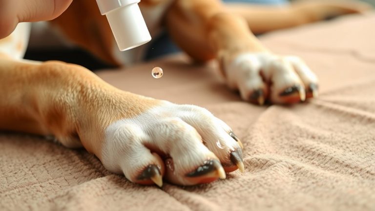
Your dog’s eye exam begins with a systematic evaluation designed to detect abnormalities before symptoms appear. The veterinarian inspects the eyelids, cornea, and anterior chamber for structural irregularities or signs of inflammation. Tear production is measured using the Schirmer tear test, a calibrated strip placed under the lower lid for 60 seconds; values below 15 mm/min indicate keratoconjunctivitis sicca. Corneal staining follows, applying fluorescein dye to reveal ulcers or abrasions under cobalt blue light. A clear, even uptake suggests surface integrity; irregular retention signals damage. Pupillary light reflexes and menace responses assess optic nerve function. Intraocular pressure is tested via tonometry, typically using the Tono-Pen Vet; readings above 25 mmHg may indicate glaucoma. A handheld slit lamp provides magnified anterior segment examination, while indirect ophthalmoscopy evaluates the retina and optic disc. Each step follows clinical protocols to guarantee diagnostic accuracy.
How Early Diagnosis Improves Treatment Success
Why do some eye conditions in dogs lead to permanent vision loss while others resolve with simple treatment? Early diagnosis makes the critical difference. You can’t always see symptoms early, but your vet can detect subtle changes during routine exams. Many dogs have a genetic predisposition to eye diseases like progressive retinal atrophy or cataracts. These are often tied to breed specific risks-Cocker Spaniels, Poodles, and Siberian Huskies face higher chances of certain disorders. Catching these conditions early allows intervention before irreversible damage occurs. Intraocular pressure, corneal integrity, and retinal function can all be measured precisely. Treatments like topical medications or surgery work best when started promptly. Delay reduces effectiveness. Regular screening turns high-risk cases into manageable ones. Early action preserves vision longer. You’re not just monitoring-you’re preventing.
Signs Your Dog Needs an Eye Exam Now
Subtle changes in your dog’s eyes can signal serious underlying conditions, and waiting for symptoms to worsen often limits treatment options. Cloudy pupils indicate possible cataracts, glaucoma, or corneal edema, all requiring prompt evaluation. Red eyes may stem from conjunctivitis, uveitis, or increased intraocular pressure above 25 mmHg, a sign of acute glaucoma. Discharge, squinting, or photophobia often accompany these signs. Unequal pupil sizes, or anisocoria, suggest neurological or ocular pathology. Vision loss, evident when your dog hesitates on stairs or bumps into objects, demands immediate attention. Corneal ulcers, detectable via fluorescein staining, progress rapidly without treatment. Intraocular pressure above 20 mmHg is abnormal in canines. Early diagnosis improves prognosis markedly. Regular monitoring using handheld tonometers and slit-lamp biomicroscopy enhances detection accuracy. These symptoms are not normal aging changes. Schedule an exam immediately if you observe cloudy pupils, red eyes, or altered vision.
How Often Should Dogs Have Eye Exams?
Regular eye exams are essential for maintaining your dog’s ocular health, especially as early detection of disease often determines treatment success. You should schedule annual exams for healthy adult dogs. However, breed predispositions increase risk for conditions like glaucoma in Cocker Spaniels or progressive retinal atrophy in Poodles, warranting biannual screening. Age factors also influence frequency-senior dogs, typically those over seven years, need exams every six months due to higher cataract and retinal degeneration rates. For geriatric dogs over ten, or breeds with known hereditary issues, your vet may recommend yearly tonometry, slit-lamp exams, and indirect ophthalmoscopy. Puppies from high-risk lines should have initial exams by six months. Monitoring intraocular pressure (normal range: 10–25 mmHg) and corneal thickness helps detect changes early. Consistent scheduling supports timely intervention.
On a final note
Regular eye exams detect subtle changes before vision loss occurs. Your dog’s corneal thickness, intraocular pressure (normal: 10–25 mmHg), and retinal function are measured precisely. Early diagnosis of conditions like glaucoma or cataracts improves treatment outcomes. For example, initiating prostaglandin analogs at the first sign of increased pressure can delay surgery. Routine screening every 6–12 months guarantees timely intervention. You preserve your dog’s vision with data-driven care.